Ruwaidix / primary research and interpretation
An image is evidence.
Not an argument by itself.
Here are four figures already present in the research archive. They are left unchanged. Open the interpretation notes to see what each source can support and what remains outside the picture.
اقرأ الصورة العلمية بوصفها دليلًا له سياق وحدود، لا نتيجة مكتملة بذاتها. جميع الصور المعروضة مأخوذة من الأرشيف البحثي المنشور على الموقع دون تعديل.
Scientific figures answer specific questions under specific experimental conditions. A photomicrograph has a different evidentiary status from a schematic; an in-vitro readout does not become a clinical outcome; and a spectrum is meaningful only with its methods and controls. The published figures are not interchangeable with raw data. For the full experimental context, follow the primary-source links below.
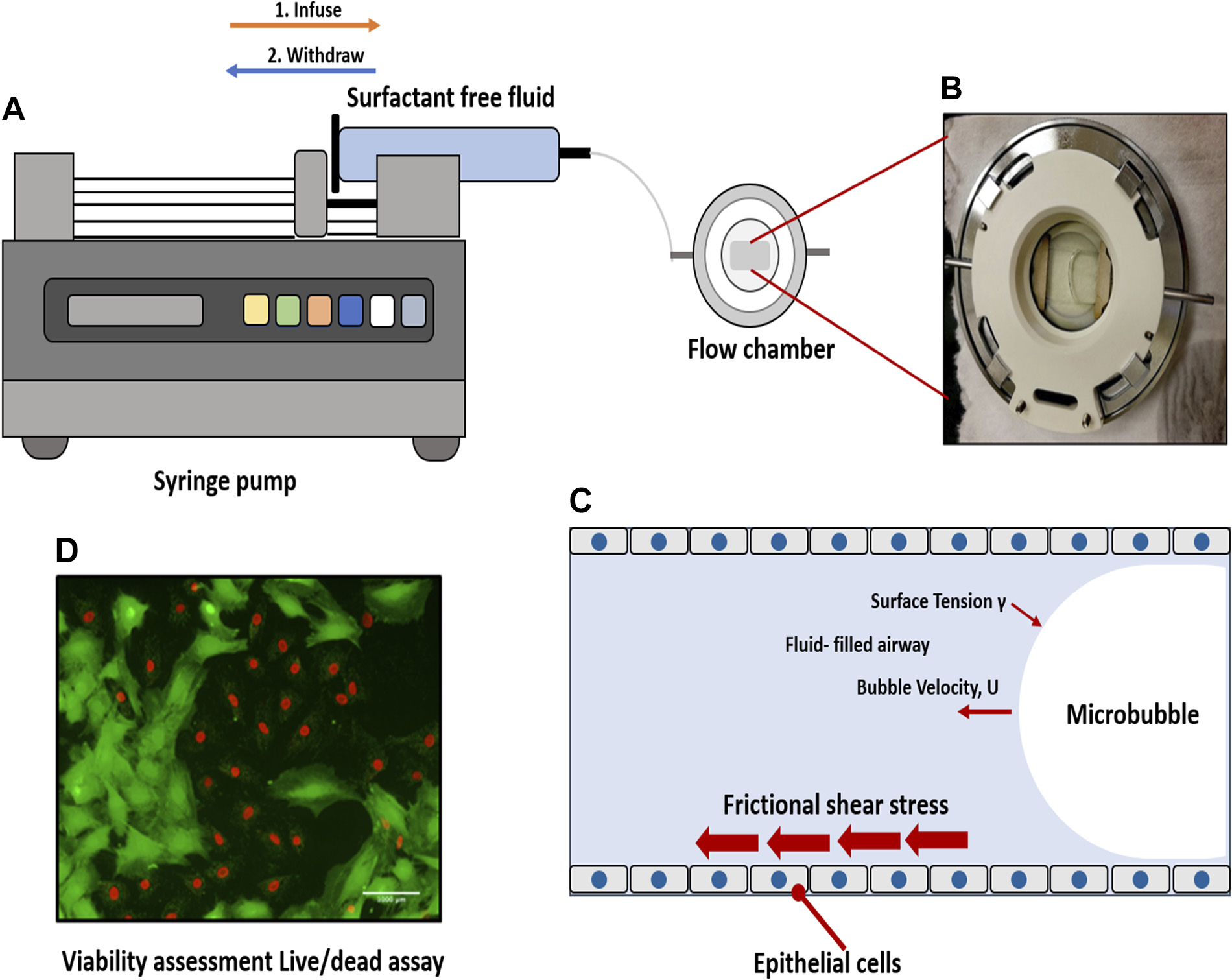
01 / Pulmonary mechanobiology
Peer-reviewed experimental study · 2021
The Journal of Applied Physiology paper ↗ documents cultured rat L2 epithelial cells, a parallel-plate chamber and a single propagating air–liquid interface. The exact panel and original legend for this stored archive image still require direct image-to-paper comparison.
A moving interface and an injured cell layer.
An experimental model asks how an air–liquid interface moving across an epithelial monolayer affects cellular viability under controlled conditions.
What this figure can help establish
The apparatus geometry and associated reported readouts provide context for experimentally imposed air–liquid interface exposure. Viability staining and other measurements must be interpreted with the paper's controls and quantification.
What this figure cannot establish
The image by itself cannot isolate a unique molecular mechanism, demonstrate therapeutic efficacy in patients, or substitute for an intact human airway. An in-vitro reduction in injury is not a clinical outcome.
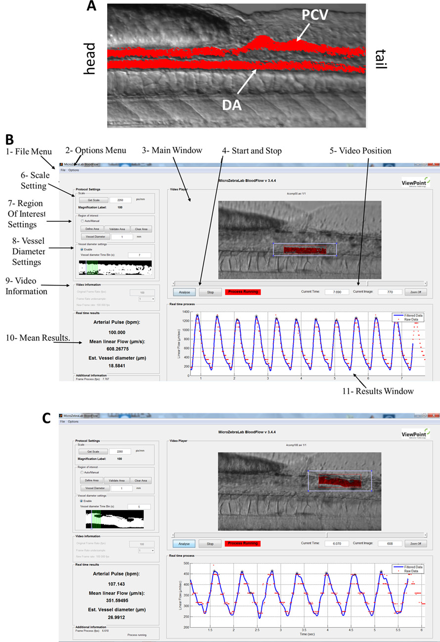
02 / Developing vessels
Peer-reviewed developmental study · 2018
The ATVB paper ↗ is the research source for endothelial cilia in developing cerebral vessels. This stored microscopy extract is not identified as an image from the separate Ramadan memory; panel-level correspondence to the paper remains to be checked.
A vessel develops inside an organism.
Developing cerebral vessels are influenced by biological context and hemodynamic conditions. Endothelial cilia and the flow environment are part of the question.
What this figure can help establish
Within the study's staged, imaged zebrafish specimens, anatomical and phenotypic observations can be related to the reported experimental groups, imaging conditions and perturbations.
What this figure cannot establish
A single microscopy image cannot demonstrate causal contributions from blood flow or cilia without the associated perturbation evidence, nor can a developmental phenotype be directly generalized to adult human disease.
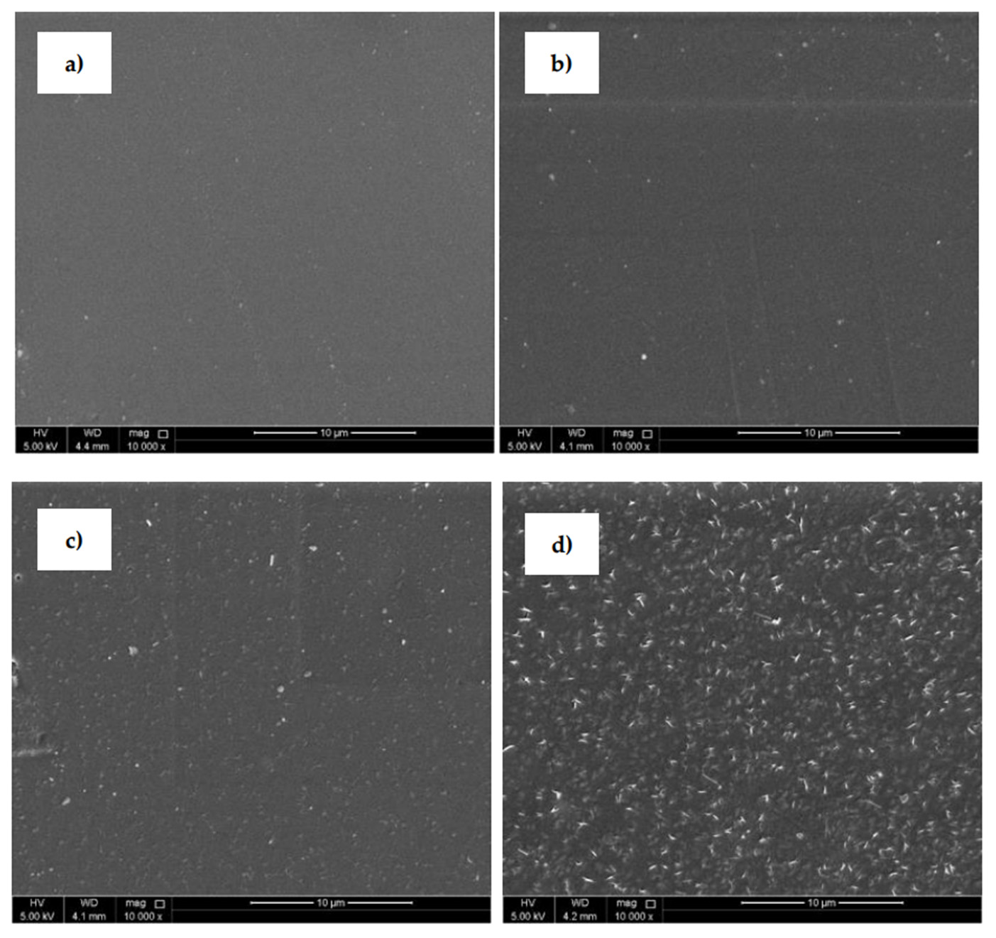
03 / Polymer interfaces
Peer-reviewed materials study · 2021
The Polymers article ↗ reports SEM comparisons of untreated, plasma-treated, ascorbic-acid-grafted and fumaric-acid-grafted PLA in Figure 5. The exact local-image panel match is not verified; consult the published figure and full legend before treating this archived extract as a particular panel.
The surface is part of the material.
Scanning electron microscopy provides morphological observations that help characterize a manufactured or treated polymer interface.
What this figure can help establish
Under the reported acquisition conditions, the micrograph may show morphological differences associated with sample processing or surface modification, subject to representative sampling and proper scale interpretation.
What this figure cannot establish
Appearance alone does not establish chemical composition, mechanical performance, antimicrobial efficacy or biocompatibility. Those claims require corresponding measurements and appropriate controls in the primary study.
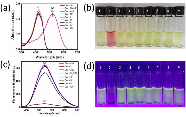
04 / Molecular sensing
Archived sensing image · source assignment pending
This file is retained from the site's imine/Schiff-base UREP23 project record ↗. Its image-to-publication attribution remains unverified. A separate curcumin cyanide-sensing paper (2019) ↗ also exists, but the shared analyte does not establish that this stored image belongs to that publication. The figure must not be presented as a verified panel from either project until its original legend is compared.
An optical response needs a chemical explanation.
A colour or absorption change is an observation under stated solution conditions. The chemistry becomes a stronger argument when spectral assignment, comparison experiments and analytical limits are included.
What this figure can help establish
With the experimental methods and controls, the spectral and colourimetric responses can support the reported sensing behaviour of the investigated imine compounds. Interpretation must follow the particular solvent, analyte and concentration conditions.
What this figure cannot establish
The figure does not by itself prove a unique binding mechanism or imply equivalent selectivity in every biological or environmental matrix. Nor does the optical response mean the initially proposed imidazopyridine synthesis succeeded.
The provenance standard
Keep the figure. Keep the context.
This viewer introduces a reading layer, not a replacement figure. Every displayed image retains its source bytes and proportions; no experimental pixels are recoloured or removed. Captions identify evidence boundaries, while original figure legends, scientific methods and publication rights must be checked in the primary sources for subsequent republication.
The publication links verify the cited papers, not automatically the identity, completeness, scale bars, resolution or reuse permissions of these local image files. Original panel legends and permissions remain a separate release check; no new figure attribution is inferred from similar-looking research subjects.
Open the complete original figure archive ↗ · Explore the documentary collection ↗