RESEARCH DOSSIER / EVIDENCE-LED RECORD
Research question, methods and contribution
Zebrafish cranial-vessel imaging, endothelial cilia, and the biological consequences of hemodynamic forces.
The scientific question
To understand how mechanical forces and endothelial cilia contribute to vascular development in the brain.
Approach and methods
Transgenic zebrafish lines, embryo imaging, flow perturbations, high-speed video microscopy, vessel-flow assessment and phenotype analysis.
PUBLISHED REFERENCE PLATES / DEVELOPMENTAL BIOLOGY
Reading an embryo before reading its vessels
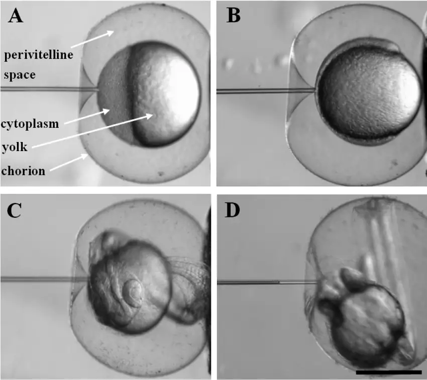
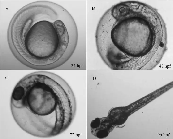
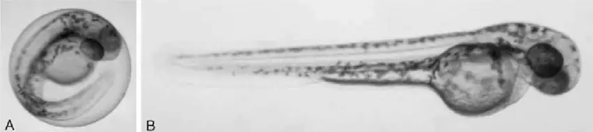
The age of a zebrafish embryo changes what a microscopist can reasonably see. These three published image plates show the anatomy and handling conditions behind developmental staging: the chorion and its changing mechanics, the visible progression from 24 to 96 hours post-fertilisation (hpf), and the difference made by removing the chorion. The images are from other research groups and are presented as contextual references, not as photographs from my experiments.
MEASUREMENT / INTERPRETATION
How the evidence is read
Embryonic vessel imaging and flow-dependent phenotypes
Developmental imaging makes vascular anatomy and endothelial-cilia distribution observable in staged zebrafish embryos. Perturbation experiments and flow measurements can help relate the observed vascular phenotypes to the conditions reported in the published study.
A developmental phenotype cannot simply be extrapolated to adult human vascular disease. The published microscopy record also cannot date or independently verify the separate Ramadan recollection associated with the work.
My documented contribution
Undergraduate member of the collaborative QU–Medical College of Wisconsin research program. Contributed experimental zebrafish imaging and practical laboratory work as part of a collaborative study. My contribution to this publication was experimental only: I did not undertake the paper's data analysis or interpretation and did not contribute to manuscript writing.
Evidence, status and limitations
The group’s work was published in Arteriosclerosis, Thrombosis, and Vascular Biology and later featured in Qatar University Research Magazine (May 2019).
Conceptual depiction of this vascular-cilia study; the microscopy references shown elsewhere on this page are from the separate publications credited above.