01 / STORIESPersonal photograph · the home microscopeصورة شخصية · المجهر المنزلي
Beginnings and questions
Early curiosity, molecular models and the instruments that made independent observation possible.
RUWAIDIX / PERSONAL SCIENTIFIC HISTORIES
In 2009 I organized a school chemistry exhibition. Two years later I was examining a fox specimen in anatomy class, struck by the difference between the body on the table and the one drawn in our textbook. University brought different objects into view: zebrafish vessels under a microscope, polymers that resisted the procedure, and a histology block that came apart before I could cut a useful section. Those experiences belong to the story as much as the papers do.
There are photographs of the microscope on my desk, colleagues between experiments, and preparations that failed. They remind me of what a published figure cannot show.
Begin with the anatomy room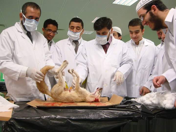
READING ROUTES / COMPLETE STORIES
Start with the school, follow an experiment into the laboratory, or meet the people who stayed in the conversation.
Early curiosity, molecular models and the instruments that made independent observation possible.
Procedures, materials and apparatus that exposed the limits of an expected result.
What microscopy and pulmonary experiments revealed about translating ideas into biological evidence.
The communities, conversations and research encounters behind the published record.
The original photographs and scientific figures appear alongside the chapters.
SCHOOL ARCHIVE / BEFORE UNIVERSITY
I organized my first school chemistry exhibition at Doha Independent School in 2009. In a photograph from a Grade 10 chemistry festival, I stand beside my first chemistry teacher, Mr. Aref Elahmad, holding the certificate I received after presenting my research. Another photograph shows my high-school graduation at sixteen, with Mr. Jameel Al-Shamarri. I remember both teachers from the years when chemistry began to occupy much of my time outside the classroom.
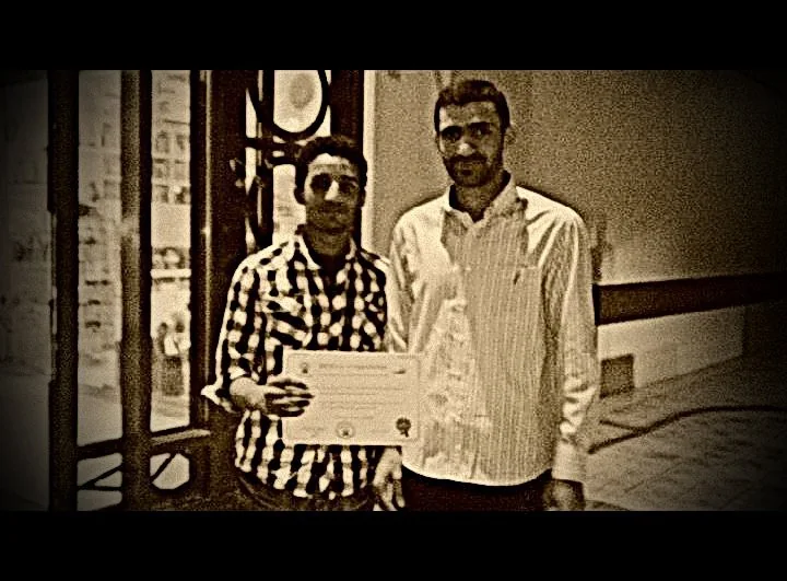
My first chemistry teacher, following my research presentation at the Grade 10 chemistry festival at Doha Independent School. The certificate in my hands belongs to that event.
مع الأستاذ عارف الأحمد، أول معلم للكيمياء درسني، بعد مشاركتي ببحث في مهرجان الكيمياء للصف العاشر بمدرسة الدوحة المستقلة. أحمل في الصورة شهادة تلك المشاركة.
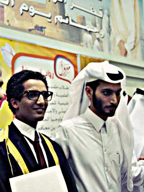
At my high-school graduation, with Mr. Jameel Al-Shamarri. This photograph belongs alongside the record of the illness that complicated my final year, but did not prevent me from graduating.
في حفل تخرجي من المدرسة الثانوية وعمري ستة عشر عاماً، مع الأستاذ جميل الشمري. لم يمنعني المرض من التخرج، وإن أثّر في امتحانات تلك السنة.
That same school period also belongs to the story of my eosinophilic illness, which continued long after graduation.
I remember the fox specimen on the anatomy table. I could examine where the organs sat in relation to one another, how much space each occupied and how different the real arrangement was from the clean boundaries of a textbook illustration. I kept trying to connect what I could see to what we had been taught in class.
I was still at school, with no experience of research, but the anatomy lesson made me pay attention to relationships between structures. Much later, the questions I encountered in vascular biology and pulmonary experiments required the same attention to the behaviour of a whole system. I could measure a component precisely and still fail to explain what happened around it.
Illness entered my life around the same period, though it followed a history of its own. The anatomy room left a different mark: direct observation, and the discovery that the tidy diagram in a textbook could not answer every question about a real body.
The photograph comes from my school archive. My account of examining a fox specimen is a memory of the anatomy lesson.
See the original account on the homepage ↗I graduated from high school at sixteen, despite spending a substantial part of my final year in hospital. My examination results kept me out of the medical school I had wanted to enter. The illness did not prevent graduation, but it changed what was possible afterwards. I eventually studied chemistry and built a life in research. I do not regard that as proof the illness was necessary or that losing medicine had been good fortune.
There were attacks when I could not keep down even water. Illness interrupted examinations and ordinary routines, while the clinical reports captured only part of what was happening. The rest belonged to the hours spent getting through each episode.
My records have used descriptions including eosinophilic gastroenteritis and hypereosinophilic syndrome. Investigations considered serious haematological disorders, later excluded. Long-term treatment brought complications of its own. These experiences shaped my educational history without defining the scientific work I went on to undertake.
When the COVID-19 lockdown began in 2020, I suffered another severe flare. Hospital access was disrupted, and concerns about the immune effects of glucocorticoids delayed my prescription. I spent fourteen days vomiting and waiting for it. Those two weeks were hell. Read the account of those two weeks ↗
In 2012 I wrote about the disease online. People continued finding me through that post years later. I now read the literature with different training, but I would still welcome a life in which eosinophils occupy less of my attention. I have other questions I want to investigate.
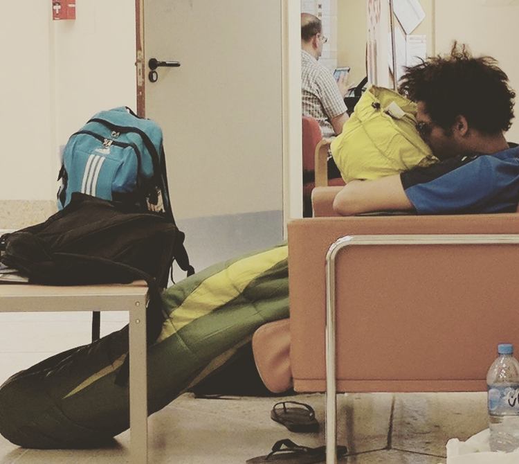
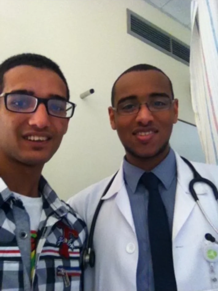
Read the longer bilingual story on this website ↗ · Read my original personal essay on Substack ↗
The personal history is based on my firsthand account in The Life Between Diagnoses and photographs I identified. Clinical terms are historical and are not offered as a definitive present-day diagnosis.
Personal account and medical context ↗University taught me techniques and equations, but much of my education took place in the hours when other students were still willing to discuss a problem after a class had ended. Helping establish the American Chemical Society student chapter at Qatar University in 2015 was part of that experience. We wanted experiments, talks and scientific conversations to reach beyond the people already working in research groups.
Hosting the fifth Chemistry Festival meant preparing explanations for visitors who could ask almost anything. I had to know where a demonstration would lead, what it could show reliably and which questions required more than we could demonstrate on the day. That preparation was as important as speaking onstage.
Helping establish the ACS student chapter meant organizing events and bringing people into the conversation. The certificate I received later reminds me of the colleagues and students who made that work possible.
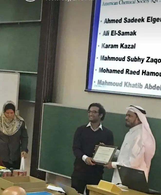
The photographs show me hosting the fifth Chemistry Festival and the certificate I received for helping establish the ACS student chapter. The chapter's activities are documented in the linked record.
Read the documented chapter history ↗Chemical synthesis had offered a pleasing degree of control. I could change a structure, adjust a condition and look for the consequence. Research on zebrafish cranial vessels and primary cilia complicated that picture. Blood flow, cell behaviour and development were not separate questions merely because we could measure them separately.
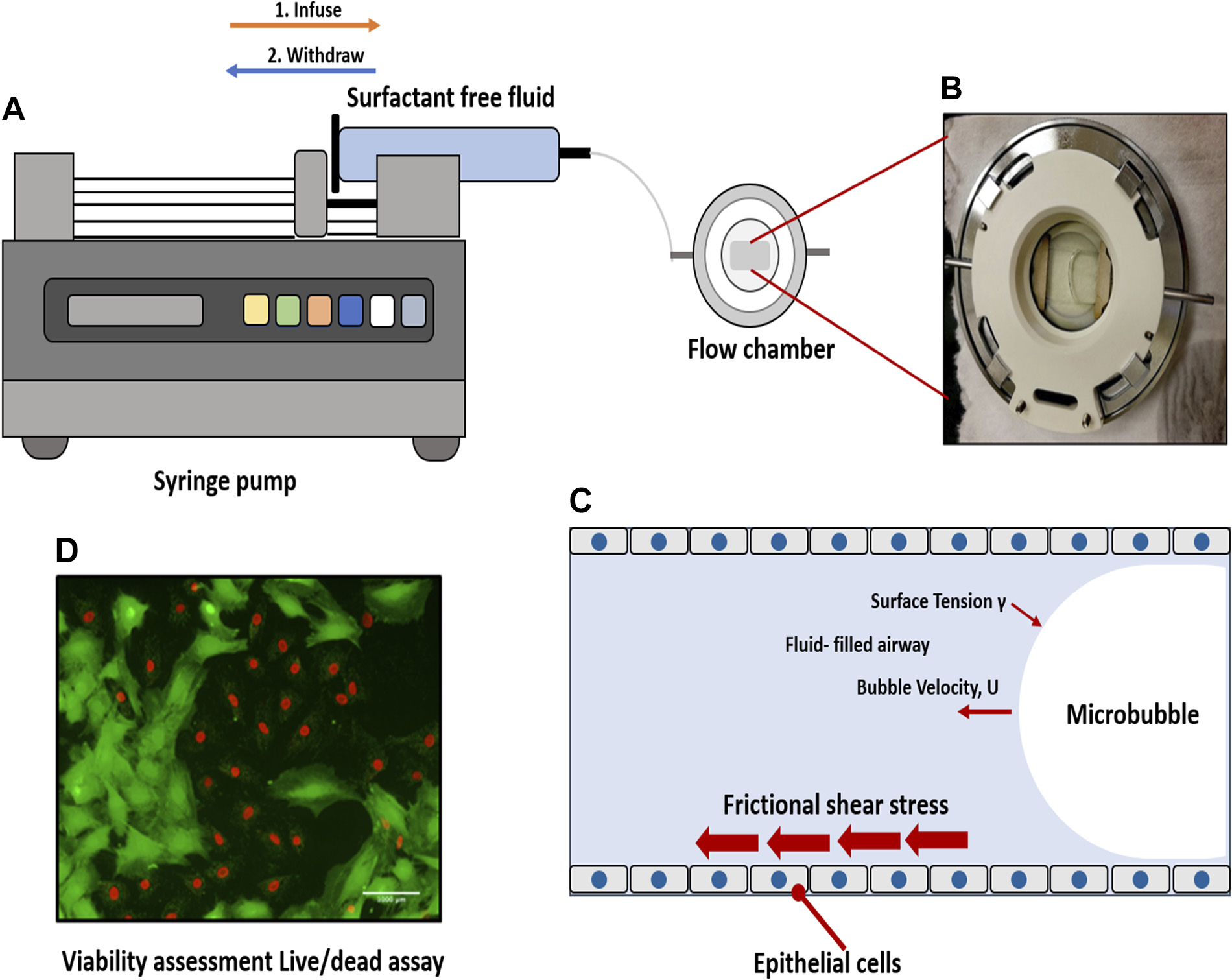
Experiments on the injury experienced by airway epithelial cells during airway reopening brought me to another scale. A force applied to a fluid could become a biological event at a cell surface. In a manuscript, the mechanics and the cellular response need to be defined and examined carefully. In the laboratory, the lesson also had a personal dimension: making a convincing model did not mean that the biology had agreed to follow it.
Among the photographs I kept are microscopes, specimen preparations and experimental apparatus. They take me back to the work before its measurements and analyses were assembled into a paper.
There is also a Ramadan memory behind this microscopy. On the final experimental day, people were waiting at my house while I was still working. Earlier evenings had meant iftar at university followed by experiments and missed Taraweeh prayers.
Read the Ramadan and ATVB story ↗The linked papers contain the methods and measurements behind the vascular and flow-chamber studies. The figures here are selected to illustrate those experiments.
Examine the experimental methods ↗Consult the figures and sources ↗On the last day of that experiment, people had already gathered inside my house. They were waiting for me, and I had not arrived. I was still at Qatar University, occupied with the work that had taken over so much of that Ramadan. I remember the contrast more clearly than I remember the hour: one room prepared for a gathering, another room where the experiment was not yet finished.
The days before had acquired a routine of their own. We would break our fast at the university, then go back to the experiments. On those evenings we missed Taraweeh prayers because we were still working. That detail matters to the memory. Ramadan is not an ordinary month with a meal shifted to sunset; its evenings have a rhythm of prayer, companionship and obligations to people. We were making choices about that time, not moving through an empty calendar.
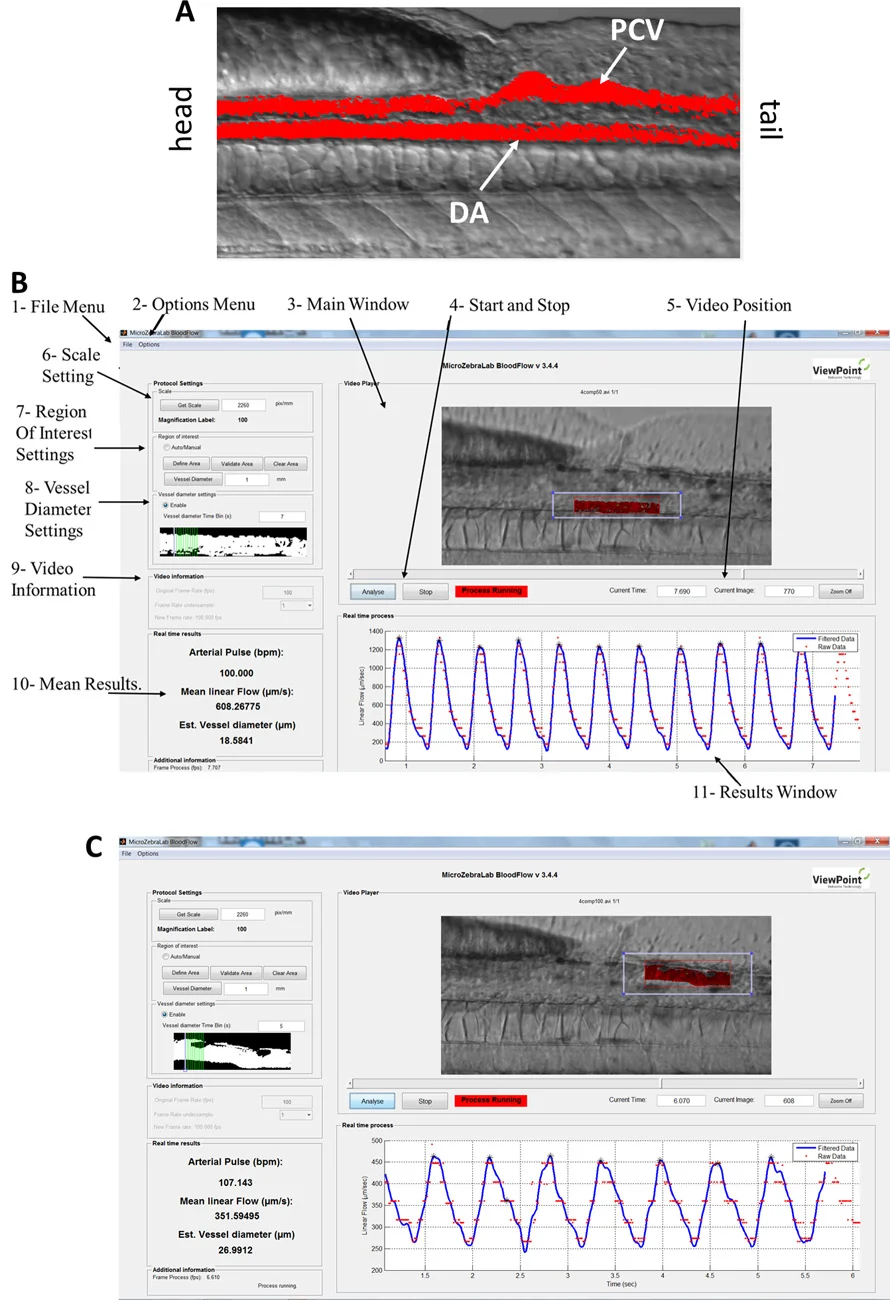
Those evenings were connected with our work on endothelial primary cilia in the developing cerebral vasculature of zebrafish. We were studying where the cilia appeared as the vascular network developed, how their distribution related to blood flow and what happened to the vessels when the cilia were disrupted. Examining such small structures required careful microscopy and equally careful interpretation of what a change in an image might mean.
On the final experimental day, I stayed at the microscope past the time I was expected home. I was reluctant to leave the session unfinished, although I knew guests were waiting for me. Earlier in Ramadan we had returned to experiments after iftar and missed Taraweeh prayers. I remember the unfinished work and the people I had kept waiting; the two occupied my thoughts throughout that evening.
The gathering at home had been planned, so my lateness affected other people. I remember wanting to finish the microscopy and knowing how much of the evening had already gone. The research eventually appeared in a paper; the hours I had taken from Ramadan gatherings and prayers were part of the experience that never appeared in its methods section.
Our collaborative study of endothelial cilia during zebrafish cerebral-vascular development was published in 2018 in Arteriosclerosis, Thrombosis, and Vascular Biology (ATVB). The figures grew out of long sessions examining the developing vessels. Reading the paper now brings back the evenings when I was absorbed in the images and reluctant to leave the microscope.
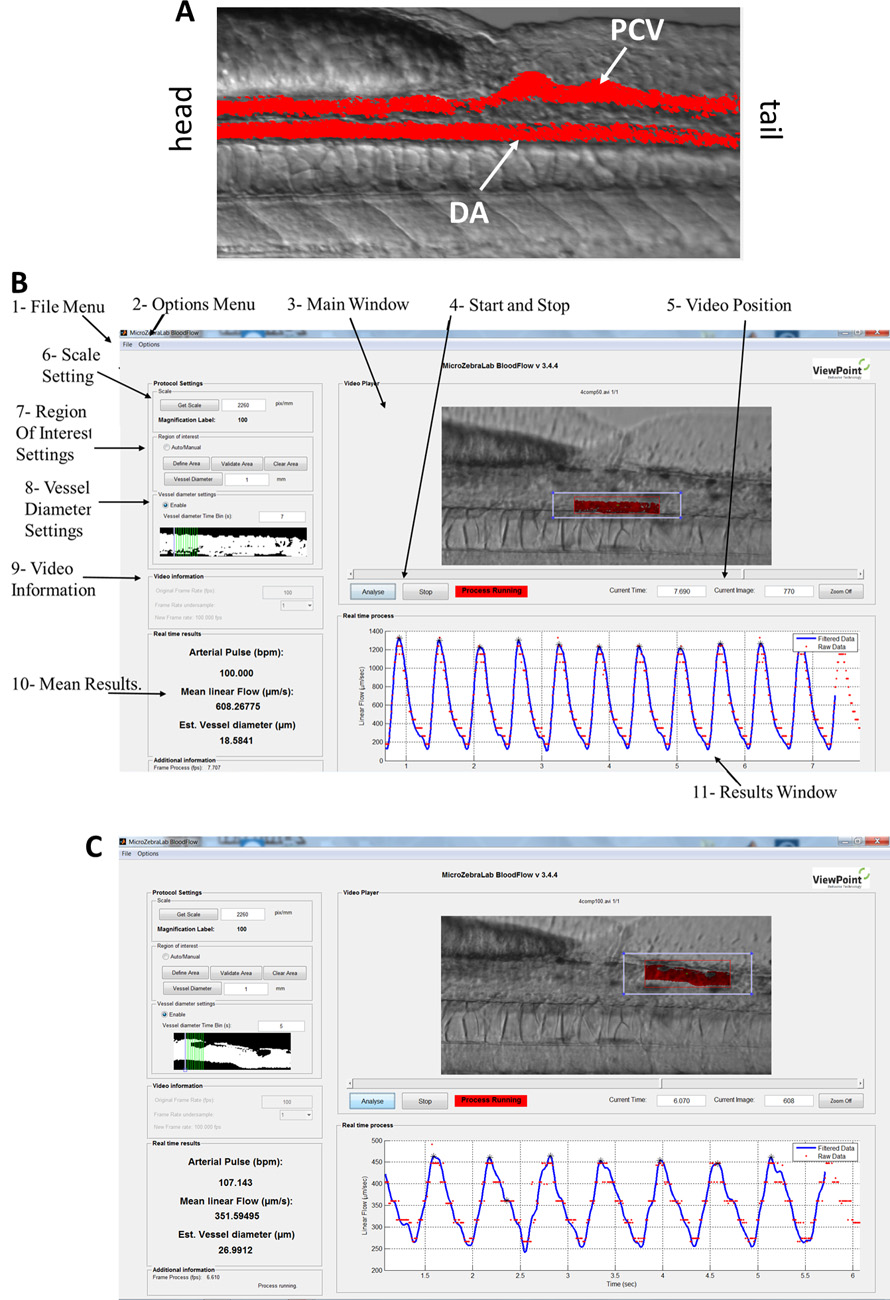
I remember breaking my fast at university, missing Taraweeh prayers on research evenings, and still being at the microscope while guests waited at my house. The photograph of the microscopy room now appears in the accompanying archive. Our 2018 ATVB paper records the zebrafish research; these evenings are my recollection of how that work fitted into Ramadan.
Read the 2018 ATVB paper ↗ Explore the zebrafish figures and methods ↗By the time I entered graduate pharmaceutical sciences in 2022, I had become interested in the distance between what a molecule can do in a controlled experiment and what it might accomplish at a site of treatment. Drug delivery is where that distance becomes difficult to ignore. A therapeutic molecule must survive the route by which we propose to administer it.
My current MSc work studies inhalable dexamethasone niosomes and proniosomes. Nebulization, aerosol behaviour and vesicle characteristics are not secondary engineering details. They determine whether a formulation's proposed advantages remain meaningful once it has to travel. After resuming graduate study in 2025, I returned to that problem with questions shaped by my earlier work on the lung.
The thesis is still in progress. Its questions concern formulation characteristics, how vesicles behave during nebulization, and what can be demonstrated about aerosol delivery. Performance and deposition results will enter the research record when their measurements and controls are complete.
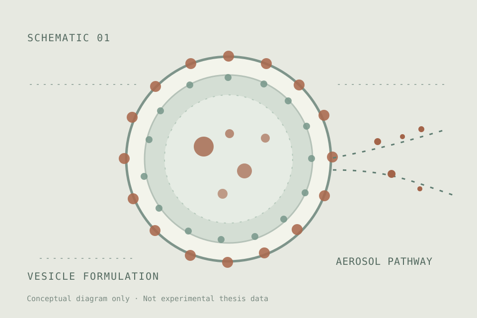
My MSc formulation work is ongoing. The illustration presents the research question; aerosol deposition and biological efficacy need their own experimental evidence.
View the current research summary ↗Read the academic record ↗OTHER SCENES / STORIES THAT DON'T FIT A STRAIGHT LINE
The next scenes move backward and forward across the years: a molecular model, community events, a microscope carried from Cairo, and a notebook filled with trials that went wrong. I returned to these memories as the research changed.
At about nineteen, I was photographed holding a ball-and-stick model of a six-membered ring. I liked being able to hold an arrangement of atoms that I was accustomed to drawing on paper. Looking at the old photograph now, I cannot tell whether the model was meant to represent benzene or cyclohexane; the bonds are not clear enough.
Later, for my undergraduate thesis on imines as chemical sensors, I worked with compounds that had to be synthesized and characterized. Each proposed structure raised questions for the spectra, and each sensing response had to be examined under controlled conditions. That was much harder than arranging the balls and sticks I enjoyed holding in this photograph.
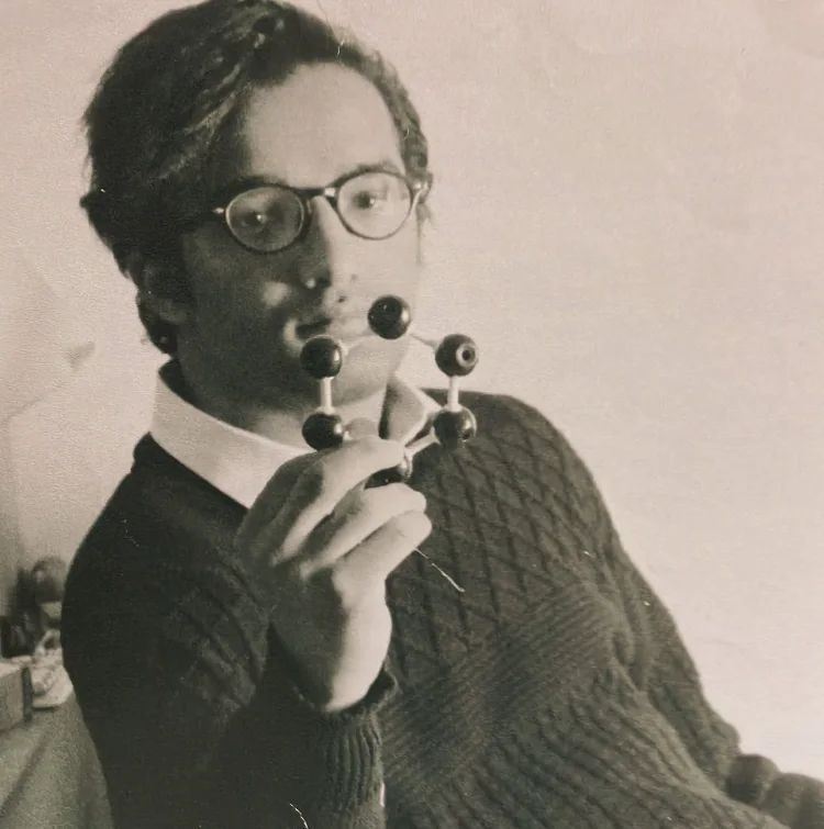
I was about nineteen when I held this molecular model for the photograph. The chemical-sensing figure nearby belongs to a separate project.
Examine the original chemistry figures ↗Some of my oldest photographs are from working with The Youth Company. I helped with breast-cancer awareness activities and the Youth Change Everything conference while I was still a student. Those events introduced me to the demands of communicating health information outside a classroom.
At those outreach events I learned that a question which seems elementary to a scientist may be urgent to the person asking it. The photographs record my participation in public-awareness work and the encounters that drew me toward more sustained scientific communication and teaching.
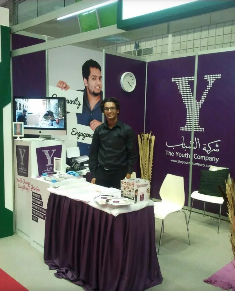
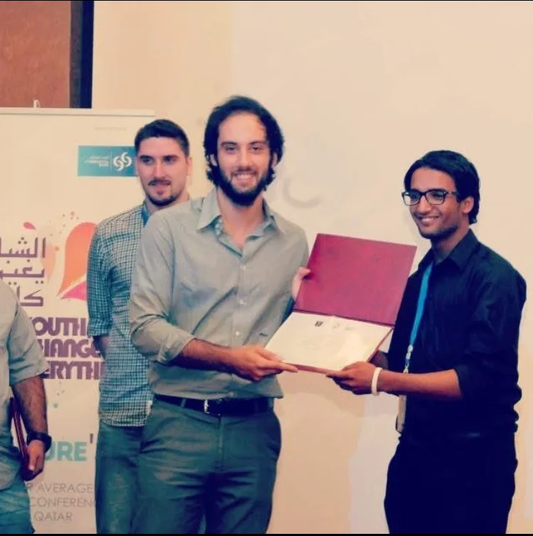
These photographs recall my activities with The Youth Company during the years before university research.
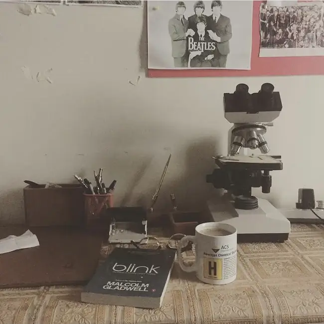
See the early-life photo archive ↗I bought this microscope in the Qasr El Einy area of Cairo, a city close to my roots, and brought it back to Qatar despite the trouble of transporting it. I wanted an instrument of my own, one I could return to whenever a question occurred to me, without waiting for the next scheduled laboratory session.
The microscope stood on my desk in Qatar among books, notes and an American Chemical Society mug. On vacation mornings I could turn to it whenever I wanted. I could spend an hour following an observation simply because I was curious, with no fixed laboratory schedule to interrupt me.
That sort of freedom mattered to my education. At university, instruments came with protocols, schedules and good reasons for both. At home, the microscope gave my curiosity somewhere to go between those structured encounters. I could follow my own interests, return to an observation and learn to notice the difference between seeing something and knowing what it meant.
I could leave a slide on the stage, return to it later and keep examining the same preparation at home. It was no substitute for the instruments and controls of a research laboratory, but having a microscope within reach changed how I spent my own time. I had gone to considerable trouble to bring it from Cairo; by 2017, it stood on my desk in Qatar, ready whenever I wanted to look at something.
I posted the photograph of the microscope at home on 1 July 2017. I had bought it in Cairo's Qasr El Einy area and brought it to Qatar, where it became part of my everyday life.
See the original photographic archive ↗Explore the research methods ↗My friends and I were working on different copolymers. Each of us had a material of our own to investigate, so even when we were sharing a laboratory and comparing notes, we were not quite performing the same experiment. Mine developed an especially stubborn problem: the polymer appeared to have become crosslinked, and it would not dissolve the way I had expected. I repeated the attempts, changed what I could, and watched the same basic difficulty return. Eventually I gave the notebook a title: A Series of Unfortunate Trials.
The solubility problem led me to think about polymer architecture. A diblock copolymer joins two different polymer segments; in a selective solvent it may form micelles, depending on the chemistry of the blocks and their compatibility with the medium. A covalently crosslinked network behaves differently because its chains cannot separate freely. Such a material may swell without dissolving. I suspected that my sample had formed a network, but failed dissolution alone could not distinguish crosslinking from aggregation, crystallinity or poor solvent compatibility.
At the time I suspected unwanted crosslinking. A failed dissolution attempt, however, cannot establish a covalent network: solvent compatibility, aggregation, crystallinity or other material changes remain possible explanations. Distinguishing them would have required systematic solvent extraction and swelling measurements alongside appropriate chemical characterization. The notebook preserves the observation and my hypothesis, not a confirmed mechanism.
What I remember most clearly is the contrast between my work and that of my friends. We had been assigned different copolymer systems, and that made the conversations about our results particularly vivid. Chemistry was no longer the tidy movement from a reaction scheme to a product. Two materials belonging to the same broad family could behave differently for reasons hidden in their molecular architecture or preparation. The experiment that would not cooperate demanded more thinking, not less.
I wrote “A Series of Unfortunate Trials” on the notebook after several attempts to dissolve the material had failed. The title was a joke about repeated failures, but I still needed to record the attempts and work out what to test next. I did not identify a crosslinked structure from those attempts. What I had was an insoluble sample, an explanation I suspected, and more characterization to do.
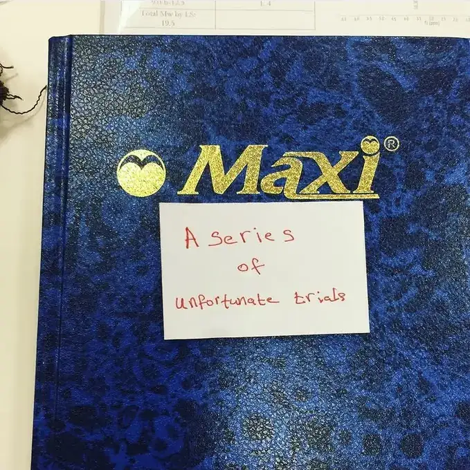
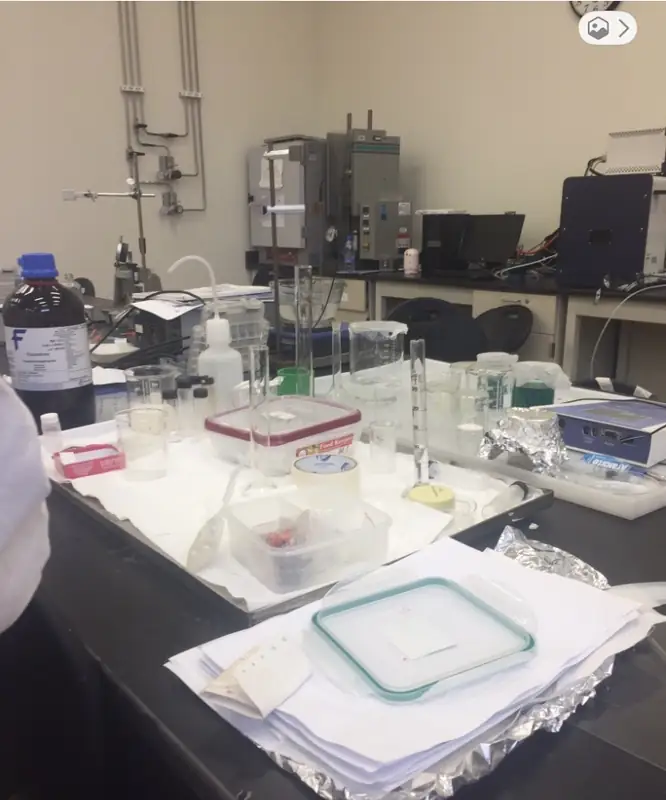
The assignment of different copolymers to friends, the difficulty dissolving my material and the interpretation of possible crosslinking are my firsthand recollections. The 8 October 2017 date is the archived post date, not a verified date for each attempt. Separately, the QNRF project UREP19-110-1-014 investigated polymeric micelles for removing polycyclic aromatic hydrocarbons from environmental samples, using commercially obtained block copolymers and Flory–Huggins compatibility calculations. Its final report does not document this synthesis setback, so I do not present the notebook as a result from that grant.
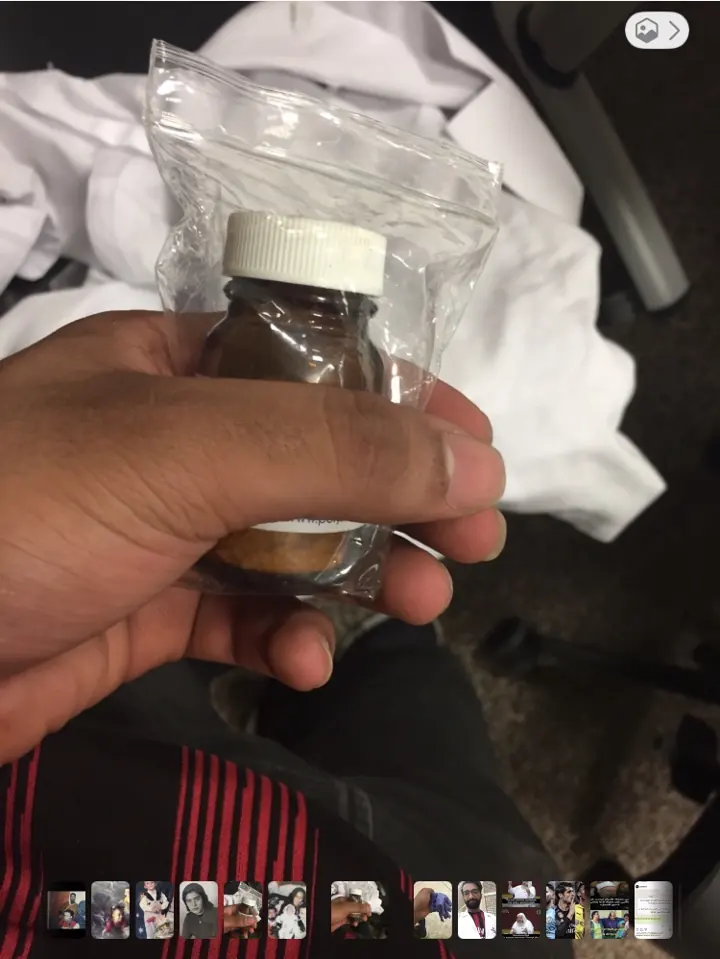
Explore the documented polymer research ↗Before a reaction became a spectrum or a figure, it began with ordinary decisions at the chemical bench, including checking what was actually in the reagent bottle. On 19 May 2018, I photographed this bottle of pyridine-4-carboxaldehyde from both sides. The two pictures record one reagent, not two different chemicals. They belong to the undergraduate chemistry work that led to my BSc thesis, Synthesis and Characterization of Imines as Chemical Sensors.
Pyridine-4-carboxaldehyde is a heteroaromatic aldehyde whose carbonyl group can undergo condensation with a primary amine to form an imine. The reverse label also records the manufacturer's assay, impurity and handling information. I kept these views as part of the material history of that work; the bottle label is not evidence of my reaction yield, the structure of any isolated product or the performance of a chemical sensor.
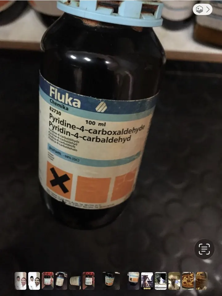
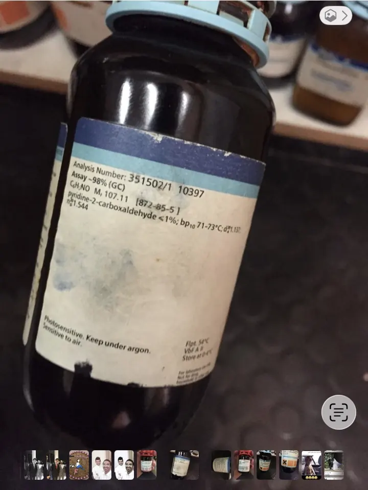
Both photographs show the same pyridine-4-carboxaldehyde reagent bottle from my undergraduate chemistry work. Its label gives CAS 872-85-5, with the supplier's information on the reverse.
See the photographic archive ↗Academic record ↗Studying history alongside chemistry changed the way I read a periodic table. A modern textbook presents its arrangement as settled; the historical record reveals the disputes, incomplete measurements and competing classifications from which it emerged. I became interested in how chemists decided which patterns deserved to be treated as laws.
A university visit involving Eric Scerri, historian and philosopher of chemistry, belonged to one side of that curiosity. An ACS student chapter astrochemistry event with Professor Yousef Hijji belonged to another. The subjects differ, but both resist the notion that a discipline is a fixed shelf of established facts. Scientific ideas have histories, and their possible applications reach into settings far beyond the laboratory we happen to know.
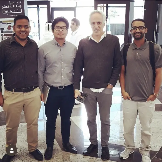
One photograph is from my university visit with Eric Scerri; another recalls an ACS astrochemistry event with Professor Yousef Hijji.
Explore the engagement chronology ↗My work across polymeric materials, electrospinning and plasma surface modification made one distinction increasingly important: the bulk chemistry of a material is not necessarily the chemistry that the cell, the liquid or the surrounding environment encounters. Fibres change geometry and accessible area. Plasma processing can change the outermost surface chemistry. Neither technique can be understood from its name alone; preparation conditions and characterization decide what was actually produced.
The archived scanning-electron microscopy figure of PLA morphology shown below is part of that research world. It invites close looking but not conclusions unsupported by its original controls. I see a continuity between this work and drug delivery, although a modified polymer surface is not an inhaled vesicle. In both, the material eventually has to meet something more complicated than the assumptions under which it was designed.
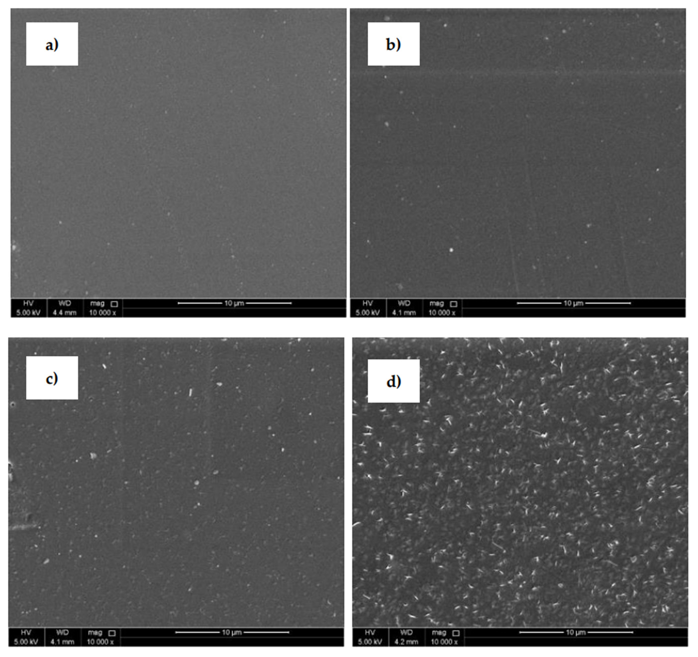
This is an archived scanning-electron micrograph of PLA morphology. The original study provides the preparation conditions and material characterization needed to interpret it.
View the materials research figures ↗The photographs bring me back to very different workspaces: pipettes on a molecular-biology bench, a stereomicroscope with positioning equipment, a biological safety cabinet and, later, a cardiovascular imaging workstation. Each setting had its own preparations and its own ways of judging whether an observation was useful.
I kept a photograph of a spectral Doppler display and a brief recording of laboratory machinery running. I remember the attention given to positioning, instrument settings and calibration, long before a result could be interpreted.
The published figure is usually the last thing a reader sees. Much of my time around the instruments was spent getting to the point where there was a measurement worth examining.
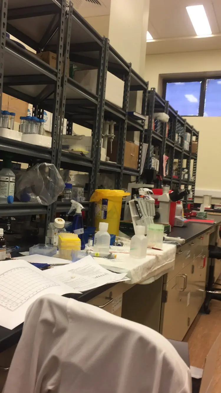
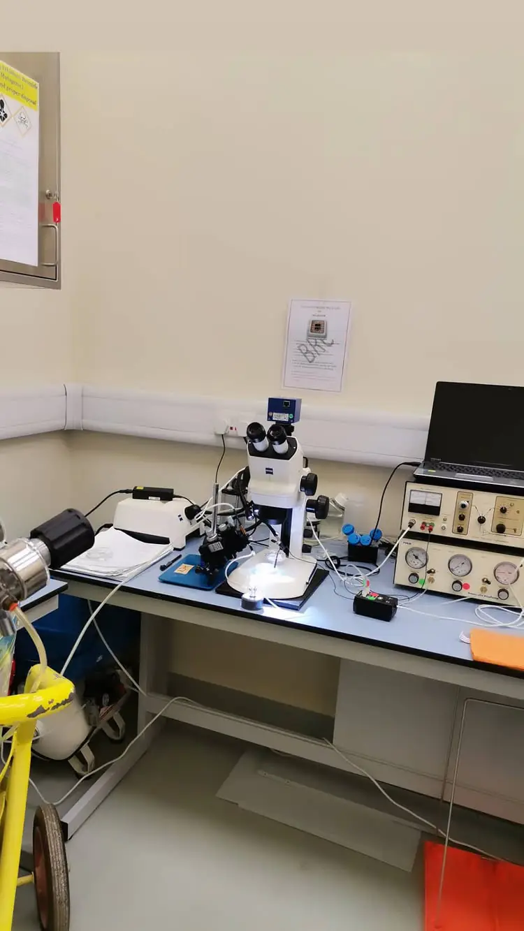
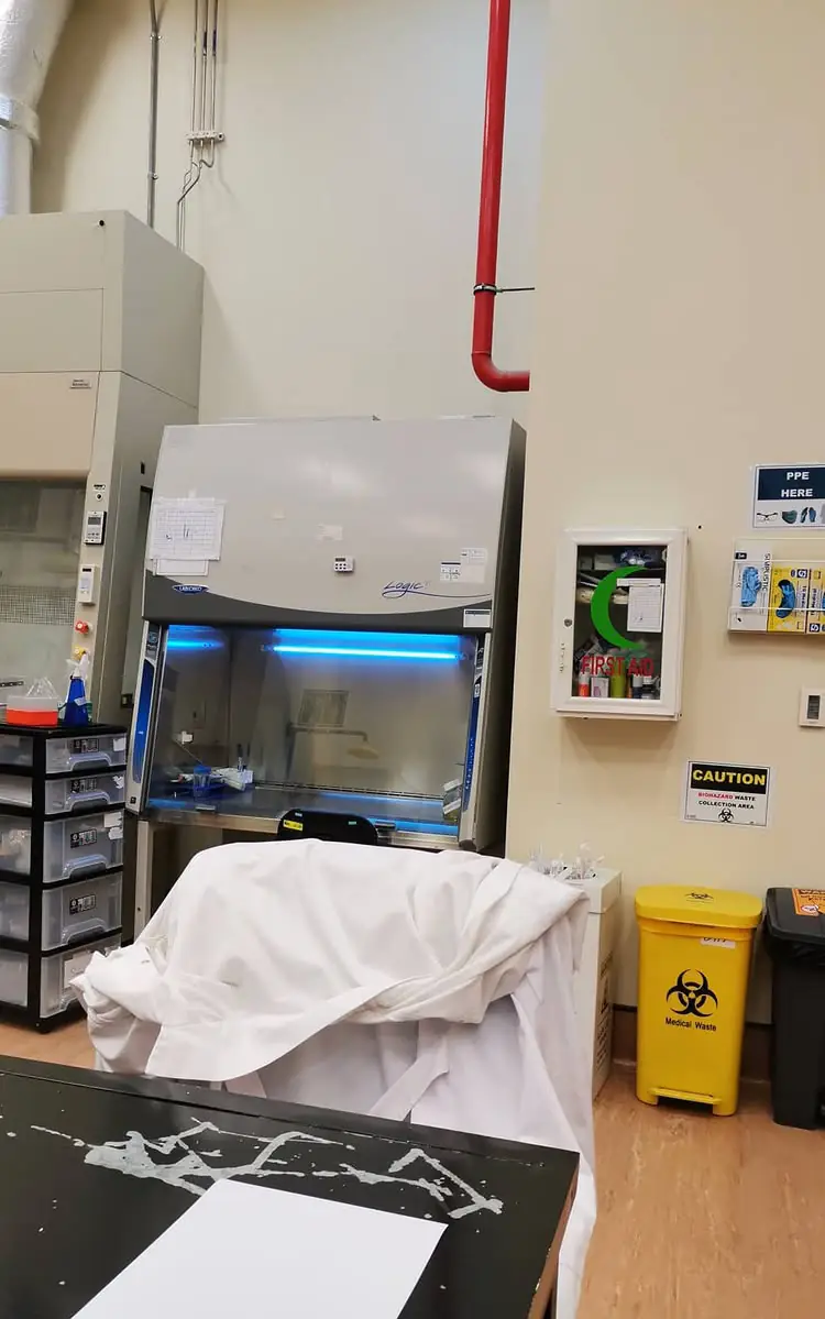
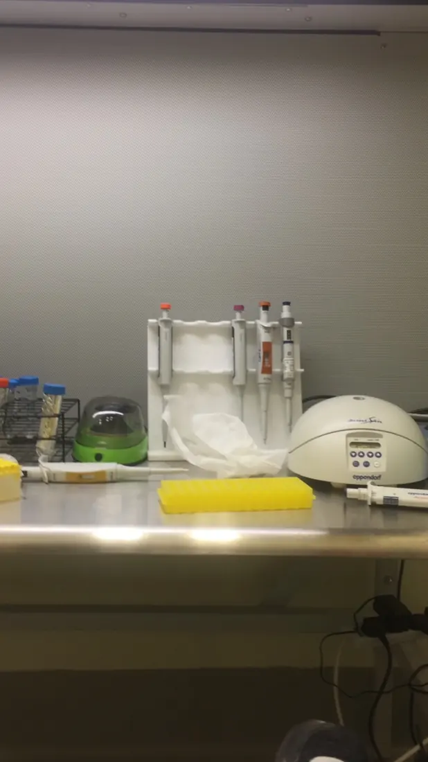
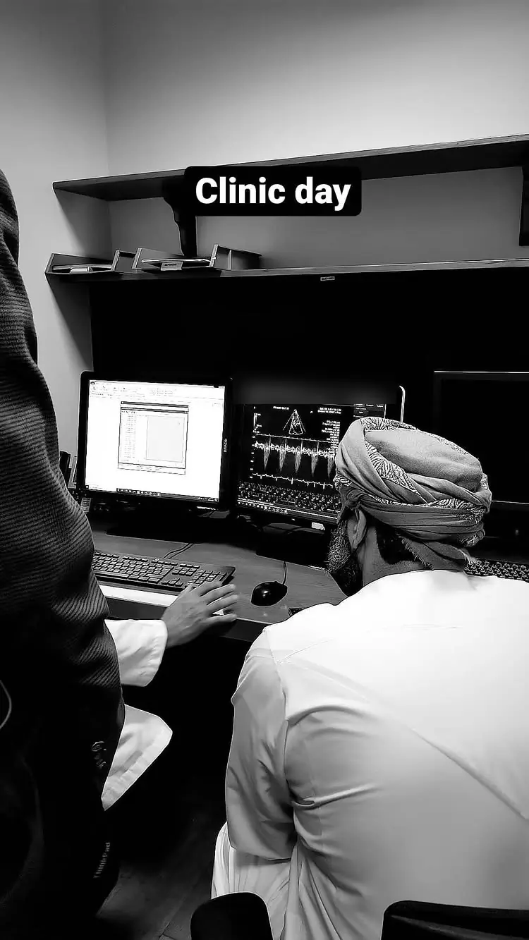
Browse original microscopy and laboratory films in Moving Evidence ↗
Separate developmental-model archive: chick-embryo incubation equipment, not part of the zebrafish endothelial-cilia experiments or the ATVB study.
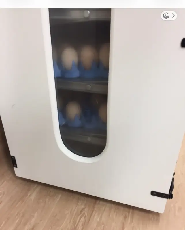
The photographs show laboratory benches, microscopy equipment and biological safety cabinets. A short recording shows an apparatus assembly in motion.
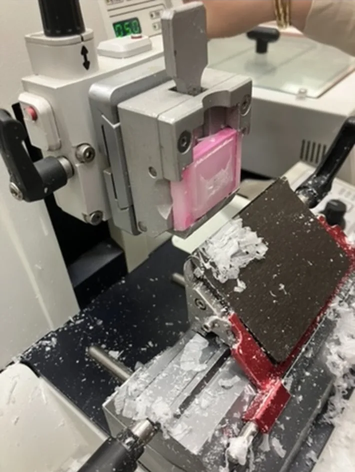
Explore the technical methods and recordings ↗I wanted to prepare serial histological sections from zebrafish embryos. The specimens were tiny, and their orientation mattered: a section through the wrong plane could miss the anatomy I was trying to examine. The practical solution I attempted was to position them first in agarose, then process that supporting block into paraffin for microtomy.
There was a reason for choosing each material. A 2% agarose matrix allowed the embryos to be held in a chosen orientation. Paraffin, following dehydration and clearing, was supposed to support thin sections. What looked like a sensible sequence turned into a problem at the interface. The agarose and paraffin formed distinguishable layers rather than one mechanically coherent block. When I attempted to section it, the material separated and broke apart. I did not obtain the reliable, interpretable sections the method required.
The microtome was not the source of the problem I could see. The agarose and paraffin had formed separate layers during processing, leaving a weak interface between a hydrophilic hydrogel and a hydrophobic embedding medium. I suspected the agarose grade and thermal behaviour might have contributed, although the photographs and failed sections could not settle the mechanism.
I kept the photographs because the attempt had failed. They show the distance between an elegant sample-preparation idea and a specimen that can actually be cut. There was no successful histopathological result to report from this attempt, only a problem worth understanding.
أردتُ إعداد مقاطع نسيجية متتابعة لأجنة سمك الزرد. وكانت العينات صغيرة إلى حدّ يجعل اتجاه تثبيتها مسألة حاسمة؛ فالمقطع الذي يمرّ في مستوى غير مقصود قد لا يُظهر البنية التشريحية التي نبحث عنها. حاولتُ أولًا تثبيت الأجنة في الآغاروز (Agarose)، ثم معالجة الكتلة الحاملة لها وإدماجها في شمع البارافين (Paraffin) حتى تصبح قابلة للتقطيع.
كان لاختيار كل مادة منطقه. فالآغاروز بتركيز ٢٪ يساعد على تثبيت اتجاه الأجنة، بينما يُفترض أن يوفّر البارافين، بعد التجفيف والترويق، الصلابة اللازمة لإنتاج مقاطع رقيقة. غير أن المشكلة ظهرت عند التقاء المادتين. تكوّنت طبقتان واضحتان بدل كتلة واحدة متماسكة، وعند التقطيع انفصلت المادة وتفتّتت. لم أحصل على مقاطع منتظمة تصلح للفحص النسيجي.
لم تكن المسألة عطلًا في جهاز التقطيع. كانت مسألة مواد وسلوكها أثناء التحضير، ولا سيما الانتقال من هلام محبّ للماء إلى وسط تضمين كاره للماء. اشتبهتُ في أن نوع الآغاروز وخواصه الحرارية ربما أسهما في المشكلة، لكنني لم أُثبت أنهما السبب الوحيد. كان انفصال الطبقتين أمرًا شاهدته؛ أما تفسيره الدقيق فظلّ سؤالًا يحتاج إلى اختبار.
احتفظتُ بهذه الصور لأن المحاولة لم تنجح. فهي تُظهر الفرق بين فكرة تبدو منطقية في خطوات التحضير وعينة يمكن بالفعل تقطيعها. لم تُنتج تلك المحاولة نتيجة نسيجية صالحة للنشر، لكنها كشفت مشكلة تستحق الفهم.
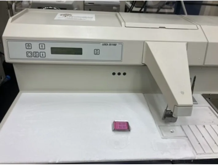
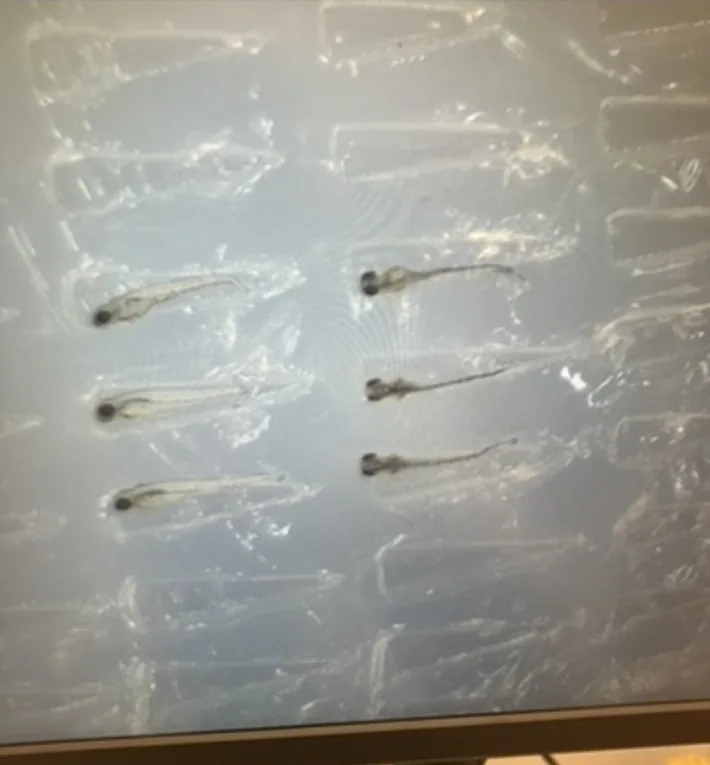
Three photographs accompany my account of the unsuccessful zebrafish embedding attempt. The archive displays 25 December 2025 for the first photograph. I observed agarose–paraffin separation; my suspicion about the contribution of agarose grade remains untested.
ترافق هذه الحكاية ثلاث صور من محاولة فاشلة لتضمين عينات سمك الزرد. يظهر تاريخ ٢٥ ديسمبر ٢٠٢٥ مع الصورة الأولى في الأرشيف. شاهدت انفصال الآغاروز عن البارافين، أما احتمال مساهمة نوع الآغاروز في المشكلة فلا يزال فرضية لم تُختبر.
I visited Zewail City in Egypt to meet Professor Ibrahim M. El-Sherbiny and explore possibilities for long-term scientific collaboration. We discussed the behavior of superparamagnetic iron oxide nanoparticles (SPIONs) and nitric oxide-releasing particles, and I learned more about the questions surrounding these systems. It was an opportunity to exchange ideas and consider where our research interests might meet.
زرتُ مدينة زويل في مصر للقاء الأستاذ الدكتور إبراهيم الشربيني وبحث آفاق تعاون علمي طويل الأمد. تناول حديثنا سلوك الجسيمات النانوية فائقة المغناطيسية من أكسيد الحديد (SPIONs) والجسيمات القادرة على إطلاق أكسيد النيتريك، وتعلّمتُ المزيد عن الأسئلة العلمية المرتبطة بهذه الأنظمة. كانت فرصة لتبادل الأفكار واستكشاف نقاط الالتقاء بين اهتماماتنا البحثية.
The second image comes from a different occasion: a portrait of me in a white coat before a wall of Arabic calligraphy. I like its place here, away from the formal portraits and experiment photographs. A person's intellectual life includes interests that cannot all be reduced to a list of techniques. I studied history alongside chemistry; the two never seemed as far apart to me as their department names suggested.
أما الصورة الثانية فمن مناسبة أخرى: صورتي بمعطف أبيض أمام جدار تزيّنه الكتابة العربية. أفضّل أن تبقى هنا بعيدًا عن الصور الرسمية وصور التجارب. فاهتمامات الإنسان لا تختزلها قائمة الأجهزة التي استعملها. درستُ التاريخ إلى جانب الكيمياء، ولم أشعر قطّ بأن المسافة بينهما تبلغ ما توحي به أسماء الأقسام الأكاديمية.
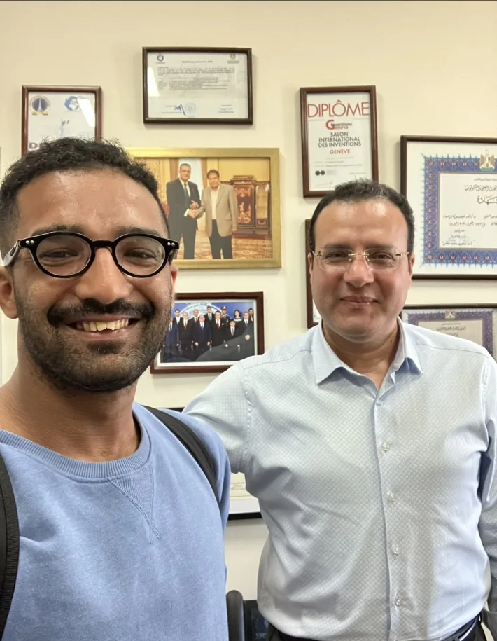
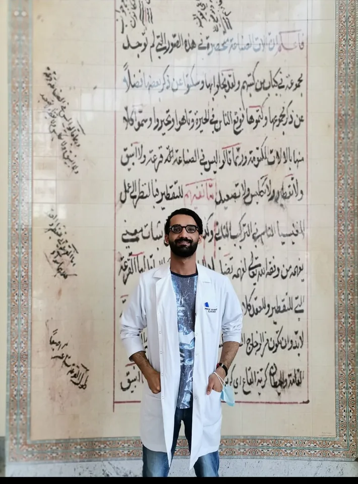
Professor Ibrahim M. El-Sherbiny, Nanoscience Program. Faculty profile linked below.
الأستاذ الدكتور إبراهيم الشربيني، برنامج علوم النانو. رابط الصفحة الأكاديمية أدناه.
Zewail City faculty listing ↗The scheme ended with an imidazopyridine. Our 2018 QNRF Undergraduate Research Experience Program proposal was built around making substituted members of that family and examining their biological activity. I joined the team as an undergraduate chemistry researcher. There were two synthesis routes in the plan, with the expected products to be characterized before biological evaluation. By the time we completed the final report in 2021, we had to record that the intended imidazopyridines had not been obtained by the proposed route.
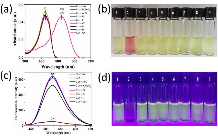
What we could isolate and investigate were imines, commonly called Schiff bases. The final report describes three such compounds and their spectroscopic characterization. They had initially occupied an intermediate position in the chemistry, but they presented questions of their own. We began examining how they responded to anions, particularly cyanide, using colour and spectroscopy instead of treating the unsuccessful ring closure as the end of the work.
One experiment gave a particularly visible answer. A yellow sensor solution shifted toward orange when cyanide was added under the conditions recorded in the report. In the UV–visible spectrum, the band near 380 nm diminished as a new band near 450 nm grew. The reported detection limit for that sensor was 1.42 × 10⁻⁵ M. Other Schiff-base compounds gave different responses to cyanide and hydroxide. Those differences mattered: a striking colour in a test tube was a useful starting observation, but it did not by itself establish selectivity or performance in an environmental sample.
The proposal had also envisaged biological evaluation. Colleagues in the biology team investigated compounds designated CMP5 and CMPB in triple-negative breast-cancer cell lines. The final report describes stronger effects of CMP5 than CMPB in several cell proliferation and migration experiments. Its measurements of SURVIVIN and c-MYC transcripts made the picture more complicated. In some treated cells, c-MYC expression increased when a reduction had been expected. Some gene-expression comparisons were not statistically significant under the reported analysis. These were in vitro experiments; they did not establish direct PRMT5 target engagement or the efficacy of a cancer treatment.
Then laboratory access became another experimental constraint. COVID-19 restrictions interrupted the later work, and the planned protein-level follow-up, including comparison with Western blot measurements, was not completed. The final document therefore contains something more complicated than an unsuccessful reaction scheme followed by a new success: it contains measurable chemical behaviour, exploratory cellular effects and mechanistic questions that were still unanswered when the project ended.
Our original proposal treated the Schiff bases as intermediates on the route to imidazopyridines. We could not complete the intended ring closure, but we isolated imines whose spectra and anion responses we could characterize. By the final report, those measurements occupied more space than the intended products. The change in direction was forced by the results: the chemistry we had actually achieved was more interesting to investigate than another account of the product we had failed to make.
The 2018 QNRF proposal and the 2021 final report describe the unsuccessful imidazopyridine route, the Schiff-base sensing experiments and exploratory cancer-cell work. Those experiments did not establish PRMT5 target engagement or therapeutic efficacy. COVID-19 restrictions interrupted protein-level follow-up.
Project aims, methods and contribution ↗Read the independent chapter ↗FURTHER READING
The notebooks, photographs and published papers lead into different parts of this work. The links below open the larger collection, including the experiments and materials that could not fit into these chapters.
Further photographs, original methods and the complete research record are linked below for anyone who wants to keep reading.