
Organisms & development
Zebrafish cardiovascular models, embryonic staging, specimen preparation and longitudinal optical observation; comparative avian embryology.
Examine methods ↗Scientific methods / The research atlas
Mahmoud Khatib A. A. Al-Ruweidi's work spans synthesis and molecular sensing, engineered interfaces, animal and cellular models, imaging, quantitative biology and drug delivery. The connecting question is how structure becomes function—and how that function can be observed, measured and changed.
A research philosophy
Chemistry establishes what a system is made of. Materials science examines how its interfaces behave. Bioengineering asks what forces, transport and architecture do to living tissue. Pharmaceutical science brings design into contact with therapeutic exposure. The history of medicine supplies a different but indispensable test: how did practitioners establish that their knowledge worked?
Methods in context
Zebrafish cardiovascular models, embryonic staging, specimen preparation and longitudinal optical observation; comparative avian embryology.
Examine methods ↗Epithelial monolayers, flow-induced injury, viability and cytoskeletal microscopy.
Examine methods ↗Electrospinning, cold plasma, polymer modification, spectroscopy and morphology.
Examine methods ↗Schiff-base chemistry, cyanide sensing, UV–Vis, fluorescence and analytical structure confirmation.
Examine methods ↗Cell-based responses, phenotypic endpoints and quantitative gene expression in collaborative research.
Examine methods ↗Video quantification, hemodynamics, biomedical data interpretation and model-informed experimental questions.
Examine methods ↗01 / Experimental imaging
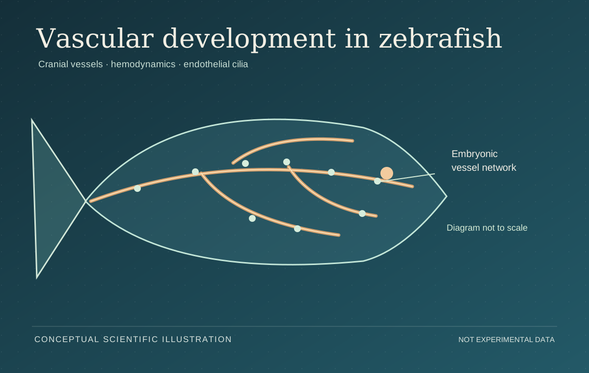
Zebrafish vascular architecture · scientific illustration.
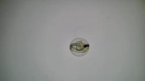
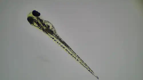
Specimen provenance, context and limitations ↗

At Qatar University’s Biomedical Research Center, Al-Ruweidi contributed to research on zebrafish cardiovascular and developmental models, linking biological preparation with high-speed optical acquisition and quantitative interpretation.
Primary visual archive / Animal methods
Recorded observation is part of the scientific evidence. High-speed zebrafish microscopy captures cardiac and vascular dynamics, while the avian laboratory archive documents an egg-preparation stage used in developmental work. The recordings below come from original files, preserving their distinct experimental contexts.
02 / Cell and tissue mechanics
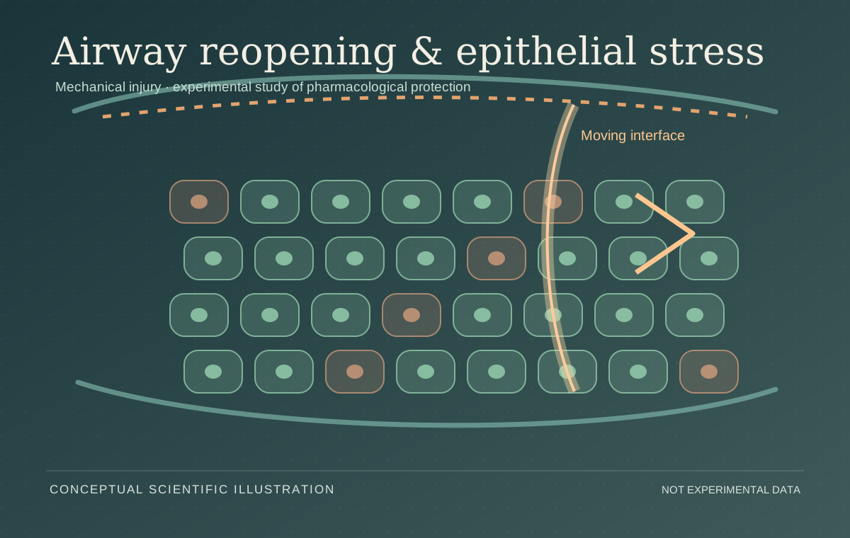
Pulmonary epithelial interface and mechanical stress · scientific illustration.

Al-Ruweidi’s pulmonary bioengineering research used an experimental model of liquid-interface-induced epithelial injury to examine how physical stress affects cell survival and cytoskeletal organization.
03 / Biomaterials · 2018–present
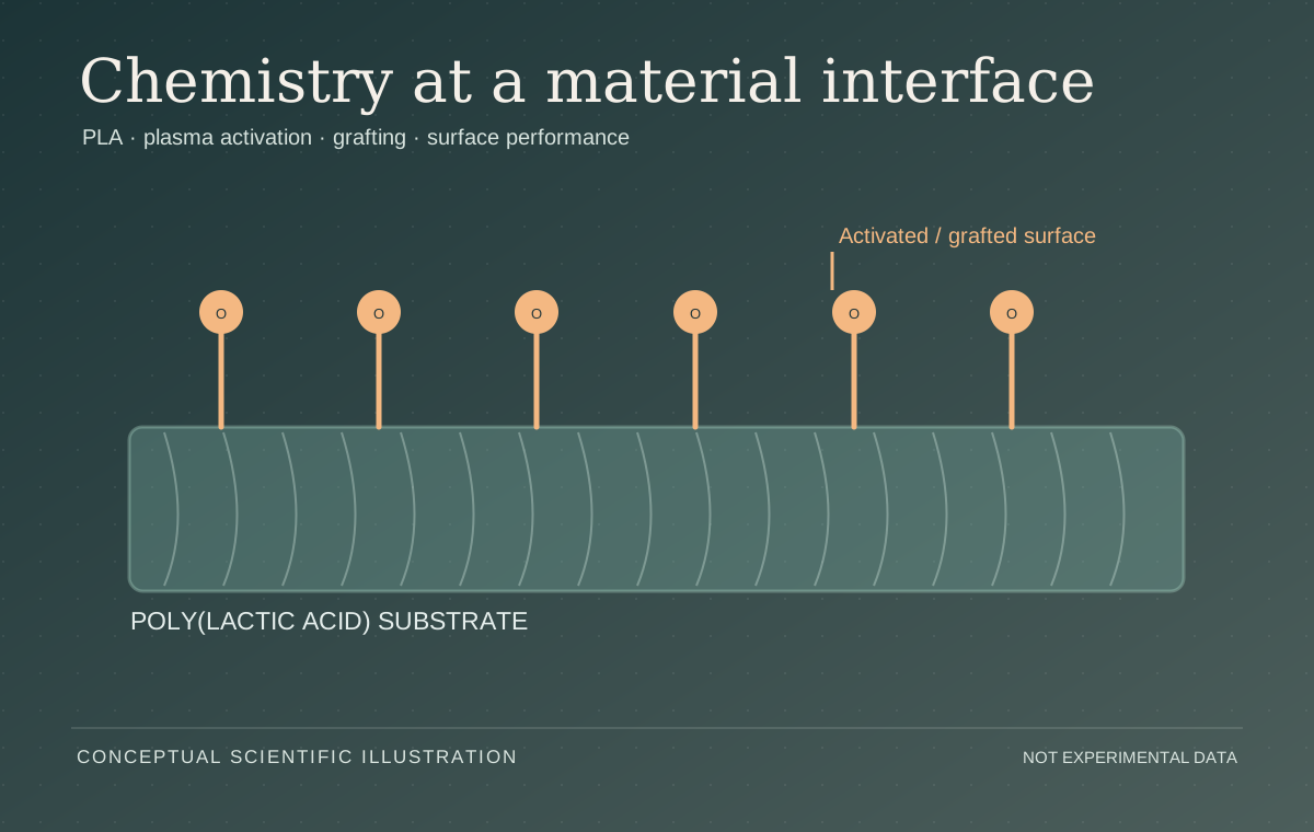
Polymeric surfaces and plasma engineering · scientific illustration.

Research at the materials–biology interface includes electrospun polymer substrates, low-temperature plasma chemistry, biomedical textiles and fabrication of functional surfaces.
04 / Pharmaceutics
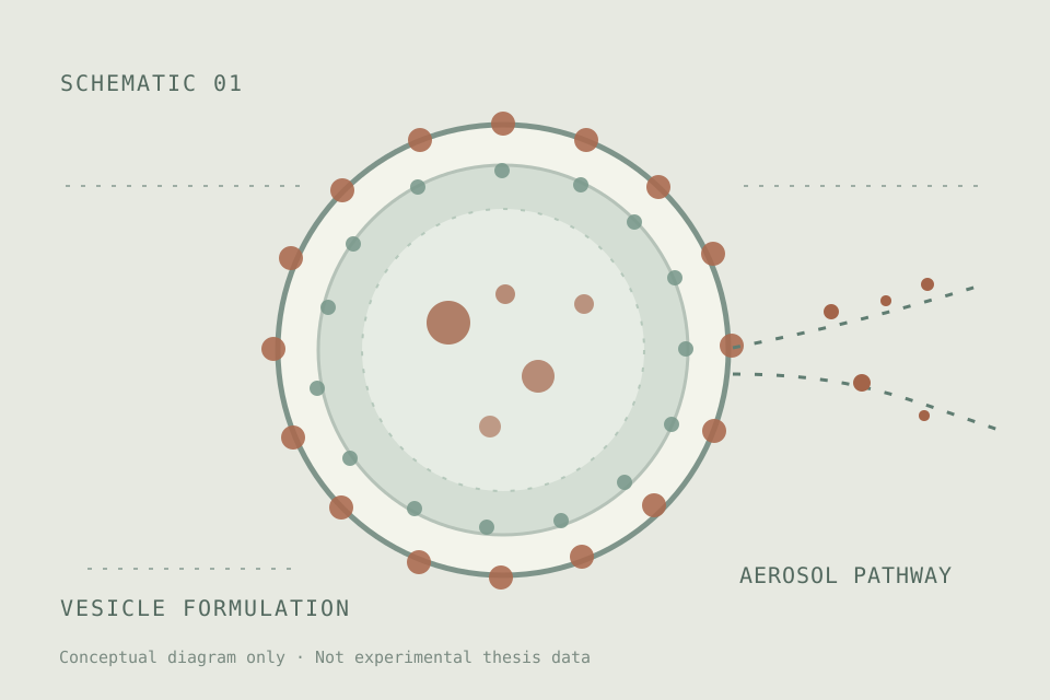
Current research · vesicular formulation and inhaled delivery.
His MSc research focuses on formulation and aerosolization questions in inhalable dexamethasone-loaded niosomes and proniosomes, bridging molecular composition, vesicular structure and pulmonary delivery.
A standard of scientific practice
Scientific techniques are strongest when the link between preparation, measurement and interpretation remains visible. Careful controls, transparent attribution and reproducible data analysis turn an isolated observation into a contribution another laboratory can evaluate and build upon.
05 / Chemistry · Synthesis · Sensor design
At the molecular scale, a measurable signal can reveal an otherwise invisible interaction.
Work in analytical and synthetic chemistry includes imine and Schiff-base preparation, chromogenic and fluorogenic probe chemistry, spectrophotometric assay design, and the interpretation of structure–response relationships. This chemical foundation connects to later investigations of polymers, therapeutics and biological materials.
Published analytical methods ↗
06 / Cell culture · Cancer biology · Biomarkers
An altered phenotype is evidence; the mechanism explaining it remains a question.
Cellular research extends from lung epithelial monolayers to collaborative work evaluating novel chemical scaffolds in cancer-cell systems. The UREP23-133-1-028 collaboration investigated Schiff-base intermediates with triple-negative breast cancer lines, including HCC1937 and MDA-MB-231, connecting molecular candidates to migration, viability and gene-expression readouts. Chemistry and biological assay teams collaborated to connect the synthesis framework to phenotypic data.
From chemical structure to cellular phenotype
Each arrow is an experimental claim that must be tested. An observed response may motivate a biomarker hypothesis, but it does not by itself establish target engagement, pathway specificity or clinical efficacy.
Related funded research context ↗07 / Structural interpretation · Characterization
A formulation, scaffold or candidate molecule is only as well understood as the evidence supporting its identity.
The characterization portfolio encompasses IR/FTIR interpretation, NMR and mass-spectrometric evidence in synthetic chemistry, UV–Vis and fluorescence behavior of molecular probes, and SEM-based surface morphology in polymer research. Molecular-structure reasoning also informs the interpretation of biomolecular interactions and the critical analysis of proposed therapeutic targets.
Published structural and morphology evidence ↗An inference chain
Those questions guide the interpretation of spectra, microscopy and composition data before biological performance is attributed to a proposed mechanism.
08 / Hemodynamics · Quantitative biology · Computation
Bioengineering connects observed dynamics to parameters with physiological meaning.
The zebrafish imaging publications use temporal microscopy alongside ImageJ and MicroZebraLab for cardiac and vascular evaluation, including heart rate, ventricular motion and blood-flow-derived indices. Related analysis extends to biomedical imaging, the interpretation of experimental endpoints, reproducible data-processing workflows and machine learning applied to fetal echocardiograms.
Published hemodynamics methodology ↗The transformation of evidence
Temporal resolution, geometry and measurement definitions determine whether video can support biological inference. The method matters as much as the result.
BRC imaging & infrastructure ↗09 / Artificial intelligence · Research software · Data analysis
Machine learning, deterministic modelling and statistical analysis belong to different methodological families. Each deserves its own evidence and limits.
Published biomedical AI: In the 2023 IEEE Access pilot study on fetal congenital heart disease, I was an equal first author. My documented contribution includes echocardiogram data curation and preprocessing, evaluation of CNN–LSTM/stacking-based approaches, clinical interpretation and scientific reporting. This is not a claim of a clinically deployed diagnostic tool.
Scientific programming and preformulation: I developed research calculations in Python using NumPy and pandas and maintain NanoFormulationDesigner, which includes physicochemical kernels, material-property provenance, screening rules, CLI/Streamlit interfaces and unit tests. Results are model-informed decisions, not experimentally validated particle-size or bioavailability predictions.
Quantitative and biomedical analytics: Python, R, SQL and Tableau support data wrangling, statistical summaries and visualization; ImageJ/MicroZebraLab support physiological image measurements; and cardiovascular data-quality, biomarker and environmental exposure analyses support scientific interpretation. The computational drug-release chapter appears as Chapter 5, beginning at page 97, in the 2026 edited volume Digital Twin Technology in Pharmaceutical Formulation and Smart Manufacturing (Deep Science Publishing; book DOI 10.70593/978-93-7185-824-3).
Dedicated AI & scientific computing research portfolio ↗Methods directly tied to evidence
Published AI: CNN–LSTM fetal echocardiography, equal first author, IEEE Access (2023).
Published AI study ↗Research software: NanoFormulationDesigner, openly inspectable repository.
Software & test records ↗Quantitative imaging: zebrafish cardiac haemodynamics and physiologic image-based measurement.
Micron methodology ↗Published visual evidence / Living archive
A moving gallery of original figures from coauthored papers. From optical images and cell mechanics to plasma-treated polymers and molecular sensing, each visual connects a technique to a peer-reviewed research record.