
Selected primary research / Original figure panels
What the experiments
actually showed.
Published microscopy, biomedical imaging and spectroscopy from my coauthored peer-reviewed papers. These scientific figures come from the source publications rather than generic illustrations. The original papers, complete methods and figure legends are linked for reproducible interpretation.

Flow-induced pulmonary epithelial injury
Published figure depicting the parallel-plate flow-chamber system, the movement of an air–liquid interface and viability assessment.
Al-Ruweidi et al. (2021), original research figure excerpt.
Read original peer-reviewed paper ↗
Quantifying zebrafish hemodynamics
Published image-analysis figure showing video microscopy of embryonic vessels and the MicroZebraLab measurement workflow.
Benslimane et al. (2020), original research figure excerpt.
Read original peer-reviewed paper ↗
Surface-engineered PLA: scanning electron microscopy
Published Figure 5: SEM panels comparing untreated, plasma-treated, ascorbic-acid-grafted and fumaric-acid-grafted PLA film surfaces.
Abdulkareem et al. (2021), Figure 5, Polymers 13(21), 3717. CC BY 4.0.
Read original peer-reviewed paper ↗
Colorimetric and fluorimetric cyanide sensing
Published spectroscopy and solution-imaging figure displaying the visual and fluorescence responses of curcumin-based sensing conditions.
Hijji et al. (2019), original research figure excerpt.
Read original peer-reviewed paper ↗Computational images and model evidence
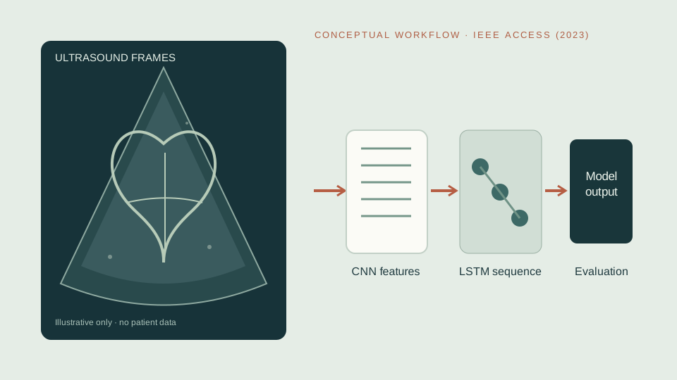
The 2023 co-first-authored IEEE Access study on fetal echocardiography is part of the research record even though the selected original-figure gallery above focuses on wet-laboratory research. Below is an original explanatory schematic, not an ultrasound video, patient image, model output or reproduced journal figure.
Peer-reviewed IEEE Access article ↗ · Contribution and methods ↗
Methods, authorship and attribution
Panels are excerpts from collaboratively authored research and are credited to their original publications. The figures represent work by the research teams credited in their respective articles. The original papers provide the complete legends, experimental methods and contribution statements. Reuse remains subject to the publisher’s applicable license; the open-access Polymers figure is published under CC BY.
Explore the techniques atlas for methodological context and the BRC imaging case study for related infrastructure.