Prepare the biological system.
Zebrafish specimens and experimental protocols relevant to embryonic development, cardiac mechanics and vessel analysis.
QU Biomedical Research Center / experimental infrastructure
On a microscope screen, a developing heart is a moving image until someone defines how to measure it. At Qatar University’s Biomedical Research Center, I contributed to establishing and developing imaging workflows for zebrafish cardiovascular research, developmental biology and experimental injury models. That work connected specimen preparation to acquisition settings and quantitative analysis so that observations could become measurements another researcher could examine.
Research infrastructure case study
I worked on preparing specimens, acquiring optical images and developing methods for quantitative analysis. That required coordination across biology, engineering and computational research, especially when equipment settings and processing steps had to be documented for other users.
Cardiac motion, vascular flow and morphological changes become evidence only when acquisition settings, temporal resolution and measurement definitions can be examined and reproduced. The infrastructure matters because the science depends upon it.
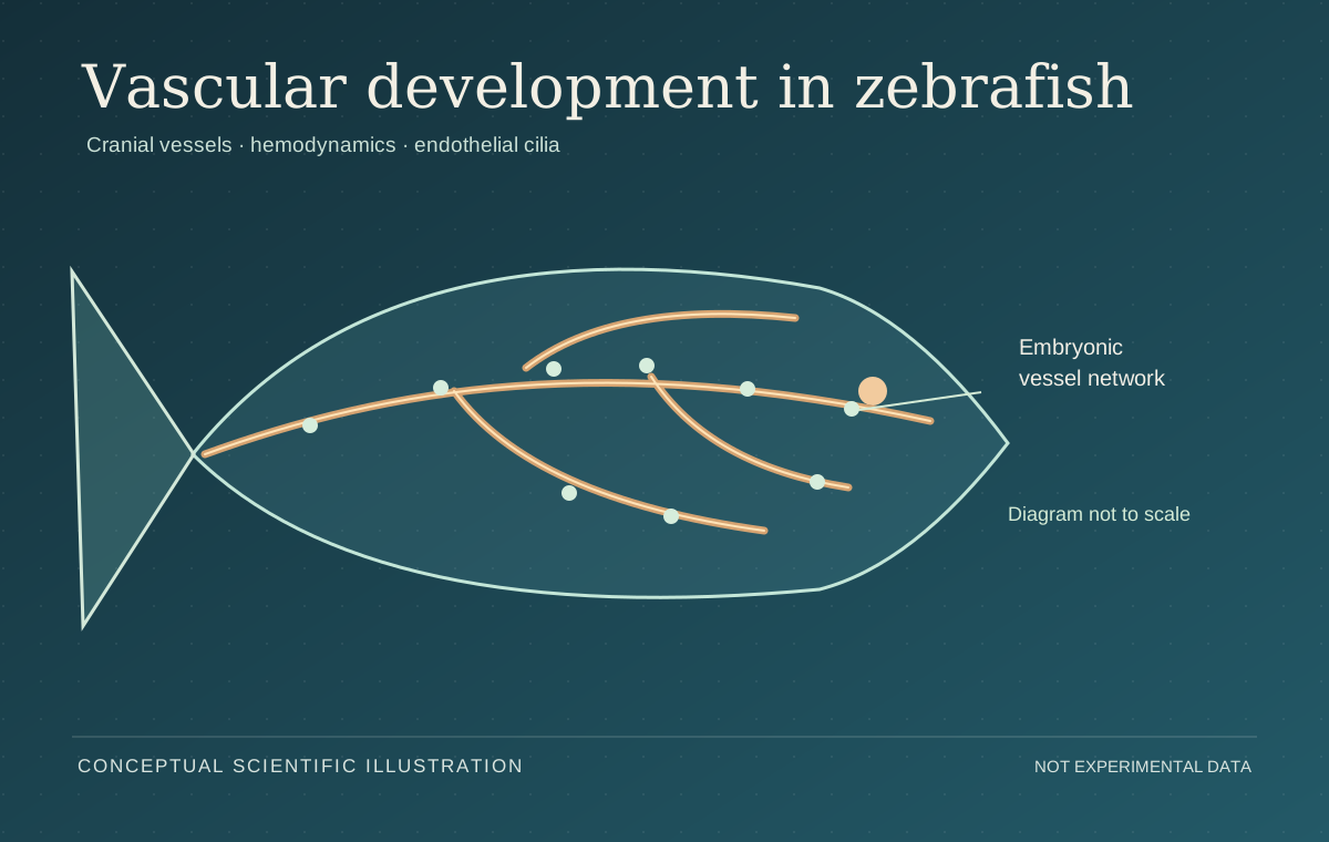
Zebrafish cardiovascular development · explanatory illustration.
The imaging workflow
Zebrafish specimens and experimental protocols relevant to embryonic development, cardiac mechanics and vessel analysis.
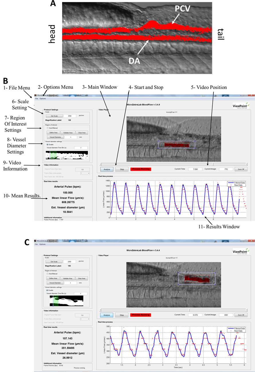
High-speed microscopy for biological processes that require temporal information, including contraction and blood-flow assessment.
ImageJ and MicroZebraLab workflows, geometric measurements, interpretation and collaboration with biologists and engineering researchers.
Protocol development, experimental preparation, reproducibility and workflow documentation within the BRC research environment.
Imaging / Mechanism / Measurement
Optical access to zebrafish cardiac function, vessel development and the mechanical environment of growing tissues. Avian embryonic systems provide a comparative context for developmental hemodynamics.
Animal models ↗High-speed microscopy followed by geometric and temporal measurements connects observable movement with cardiac and vascular parameters.
Quantitative biology ↗For the AI-driven fetal echocardiography study and its model-evaluation methods, see the published CNN–LSTM case study ↗.
Cell imaging and controlled mechanical exposure reveal biological consequences that an end-point viability number alone may conceal.
Cellular mechanobiology ↗Protocol development, instrument readiness, image documentation and training connect individual studies to an enduring research environment.
Published figure archive ↗Research collaboration / Institutional context
The zebrafish vascular-development work was one part of a wider collaboration across the two institutions. This Qatar University Research seminar discusses the opportunities and challenges of international research collaborations. The recording provides institutional context; the papers on this page remain the sources for experimental findings.
Open original seminar recording ↗Published research context
Benslimane et al., “Cardiac function and blood flow hemodynamics assessment of zebrafish using high-speed video microscopy,” Micron 136 (2020), 102876. Eisa-Beygi et al., “Characterization of endothelial cilia distribution during cerebral-vascular development in zebrafish,” Arteriosclerosis, Thrombosis, and Vascular Biology 38 (2018), 2806–2818.
Micron paper ↗ ATVB paper ↗